Pediatric Stroke: Why It's Missed for Days, the Arteriopathies, and the PedNIHSS
The average childhood stroke takes roughly a day to diagnose — not because it's subtle, but because nobody in the chain thinks "stroke" about a child. The differential is different, the scale is adapted, and the delay is the modifiable part.
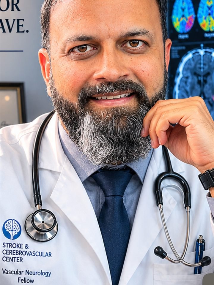
In pediatric stroke, the modifiable risk factor is the delay.
- →Childhood stroke is rare but real — and diagnosis is routinely delayed by many hours to days, because mimics are common and nobody's first thought about a child is "stroke."
- →Age changes the presentation: neonates present with seizures (the hemiparesis often appears months later as an emerging hand preference); older children present like adults — focal deficits — plus seizures and headache far more often than adults do.
- →The etiology list is its own world: arteriopathies lead (focal cerebral arteriopathy, moyamoya, dissection), then congenital heart disease, sickle cell disease, prothrombotic states, and infection-associated mechanisms.
- →MRI is the preferred study (rapid protocols where available) — a normal head CT excludes little in a child. Vessel imaging belongs in the same visit.
- →Deficits are quantified with the PedNIHSS — the validated 2–18 adaptation. Hyperacute reperfusion in children remains case-by-case at experienced centers; sickle cell disease has the field's best-proven prevention (TCD screening and transfusion).
The 9-year-old's right arm stopped working at soccer practice at 4 p.m. He was seen at an urgent care at 6, diagnosed with "probably a migraine," and sent home. He returned the next morning because the arm still hung; the MRI at hour 20 showed a left MCA-territory infarct. Nothing about the case was subtle. What was missing, at every stop, was the thought.
That is the defining fact of pediatric stroke, and the professional statements say it plainly: diagnostic delay is the rule, driven by low suspicion and mimic-rich presentations, and shortening it is the single most modifiable part of the disease.1
Presentation by age: two different diseases wearing one name
Neonates (a peak period for stroke incidence) rarely announce a deficit. Perinatal arterial ischemic stroke presents most often with focal seizures in the first days of life, or with nothing at all — discovered months later as an emerging early hand preference or asymmetric reaching. A baby with a definite hand preference before their first birthday deserves imaging, not reassurance.1
Children and adolescents present the way adults do — hemiparesis, facial droop, aphasia, visual and gaze findings — but with two loud differences: seizures at onset and headache are common companions, and the mimic list (migraine with aura, Todd paresis, functional presentations, tumor, demyelination) is genuinely longer than in adults. The operational answer is not cleverer guessing; it is a lower imaging threshold for any acute focal deficit, with the same "sudden and focal means vascular until proven otherwise" reflex we teach for adults in what is a stroke.
The etiology list children actually have
- Arteriopathies — the leading category. Focal cerebral arteriopathy (a unilateral, often post-infectious stenosis of the distal ICA/proximal MCA, classically after varicella), moyamoya (childhood ischemia provoked by crying and hyperventilation — its own chapter: moyamoya), and craniocervical dissection, often after trivial trauma (mechanism and management logic in the dissection chapter).1
- Cardioembolism — congenital heart disease, recent cardiac surgery or catheterization, mechanical support: the reason echocardiography sits high in the pediatric workup.
- Sickle cell disease — historically among the strongest stroke risks of childhood, and the field's best prevention story: transcranial Doppler screening with transfusion programs for high-velocity children dramatically reduces first stroke. It is the one pathway every system should have wired.1
- Prothrombotic states and the rest — inherited and acquired thrombophilias (more relevant here than in adult arterial stroke), infection and inflammation, genetic arteriopathies overlapping the young-adult zebra list.
Imaging and scoring: MRI first, PedNIHSS at the bedside
MRI with diffusion is the study of choice — a normal CT is close to meaningless for excluding ischemia in a child, and radiation economy matters at this age. Rapid-sequence protocols increasingly make MRI feasible in the emergency window; vessel imaging of head and neck belongs in the same session, because arteriopathy is the leading answer and it changes management.1
Quantify the deficit with the Pediatric NIHSS — the age-adapted version validated for 2–18, scored prospectively with excellent interrater reliability.2 We host a guided version with the task modifications inline: the PedNIHSS calculator.
A normal head CT in a child with a new focal deficit has excluded almost nothing. The MRI is the exam.
Treatment: honest hierarchy of evidence
Hyperacute reperfusion in children is where honesty matters most: there is no completed randomized trial (the dedicated pediatric alteplase trial closed for lack of enrollment), so thrombolysis and thrombectomy are individualized decisions at experienced centers — most defensible in adolescents who meet adult-style criteria with confirmed large-vessel occlusion, made with pediatric stroke expertise at the table.1 What generalizes from adult care is the systems thinking, not the checklist: rapid recognition, parallel imaging, and a predefined pathway.
Beyond the window, care is supportive neuroprotection (normoglycemia, normothermia, seizure control), antithrombotic therapy matched to mechanism — commonly aspirin, with anticoagulation for cardioembolic sources and dissection per institutional practice — arteriopathy-specific care (revascularization for moyamoya, immunomodulation debates in focal cerebral arteriopathy), and the sickle-cell pathways above. And because it is childhood, rehabilitation and school reintegration carry decades of leverage: plasticity is real but not a guarantee, and outcomes range from full recovery to lifelong hemiparesis, epilepsy, and learning effects that surface years later. Follow-up is a long game played with the family.
The bottom line
Children stroke differently at every step: seizures instead of deficits in neonates, mimic-rich presentations in childhood, arteriopathies and hearts instead of atherosclerosis, MRI instead of CT, an adapted scale, and therapy decisions that lean on judgment more than trials. The one constant is the choke point — thinking of it. Put pediatric stroke on the differential for any acute focal deficit, seizure with focal features, or infant hand preference, and the rest of the pathway finally gets its chance to work.
Frequently asked questions.
How do strokes present in children?
By age. Neonates most often present with focal seizures in the first days of life, or silently — appearing months later as an early hand preference. Older children present like adults (weakness, facial droop, speech or visual change) but with seizures at onset and headache far more often, which is part of why the diagnosis gets missed.
Why is pediatric stroke diagnosed so late?
Because suspicion is low and mimics are common: migraine, Todd paresis after seizure, functional symptoms, tumor. Typical series report many hours to more than a day from onset to diagnosis. The fix is a lower threshold for urgent MRI in any child with an acute focal deficit.
What causes stroke in children?
Arteriopathies lead — focal cerebral arteriopathy (often post-infectious), moyamoya, and craniocervical dissection — followed by congenital heart disease and cardiac procedures, sickle cell disease, prothrombotic states, and infection-related mechanisms. Atherosclerosis is essentially absent from the list.
What is the PedNIHSS?
The pediatric adaptation of the NIH Stroke Scale, validated for ages 2–18, which modifies tasks to be developmentally appropriate while keeping the adult structure and scoring range. It showed excellent interrater reliability in prospective validation and is the standard way to quantify deficits in childhood stroke.
Can children receive tPA or thrombectomy?
Sometimes — as individualized decisions at experienced centers, most defensibly in adolescents meeting adult-style criteria with a confirmed large-vessel occlusion. No completed randomized trial exists in children, so guidelines stop short of routine recommendations and emphasize pediatric stroke expertise in the decision.
How is stroke prevented in sickle cell disease?
With transcranial Doppler screening: children whose TCD velocities are high are at markedly elevated stroke risk, and regular transfusion programs in that group dramatically reduce first strokes — the best-proven prevention in all of pediatric stroke. Hydroxyurea has a role in maintaining risk reduction under specialist guidance.
References.
- Ferriero DM, Fullerton HJ, Bernard TJ, et al. Management of Stroke in Neonates and Children: A Scientific Statement From the American Heart Association/American Stroke Association. Stroke. 2019;50(3):e51-e96. PubMed
- Ichord RN, Bastian R, Abraham L, et al. Interrater reliability of the Pediatric National Institutes of Health Stroke Scale (PedNIHSS) in a multicenter study. Stroke. 2011;42(3):613-617. PubMed
- Scott RM, Smith ER. Moyamoya disease and moyamoya syndrome. N Engl J Med. 2009;360(12):1226-1237. PubMed
Related guides
Keep building the picture.
- PedNIHSS calculator The validated 2–18 adaptation, with task modifications inline.
- Moyamoya disease The childhood arteriopathy provoked by crying and hyperventilation.
- Cervical artery dissection Trivial-trauma dissection is a leading pediatric mechanism too.
- Stroke in young adults The adjacent age band and its overlapping zebra list.
Article written by